
I will be attending the Brain Mets 2017 Conference in Marseille, France October 6-7. More information can be found at http://brain-mets.com/
If you are there I look forward to meeting you. Abstract below:
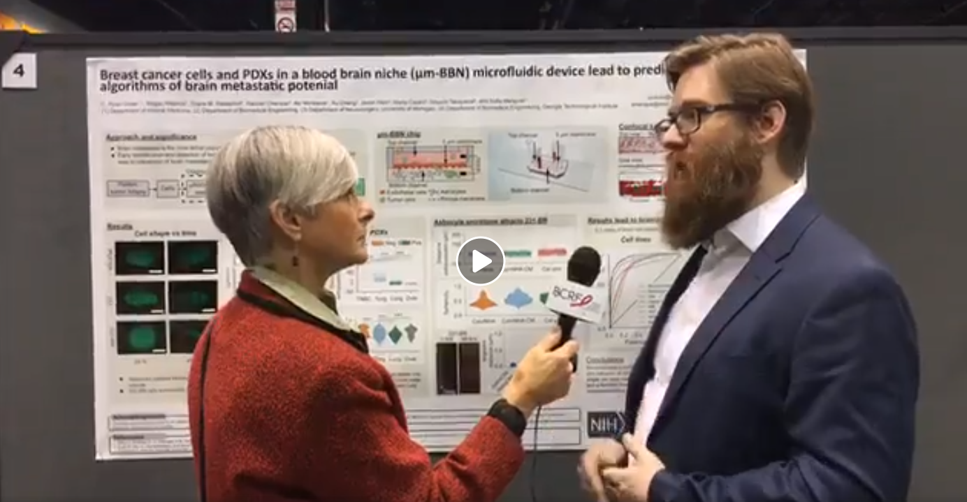
Development of a blood brain niche microfluidic device and algorithms to aid diagnosis of brain metastatic potential
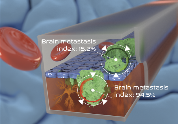
Brain metastasis is the most lethal complication from advanced cancer. 15% of breast cancers metastasize in the brain with a low one-year survival rate. A critical step in reducing the lethality of brain metastasis is early detection of clones with high metastatic potential. Models for characterizing metastatic potential include murine models and in vitro microfluidic models. Murine models are costly, time intensive, slow to manifest metastasis and difficult to analyze. On the other hand, in vitro systems are faster and cost effective but currently do not recapitulate the “live” micro-environment.
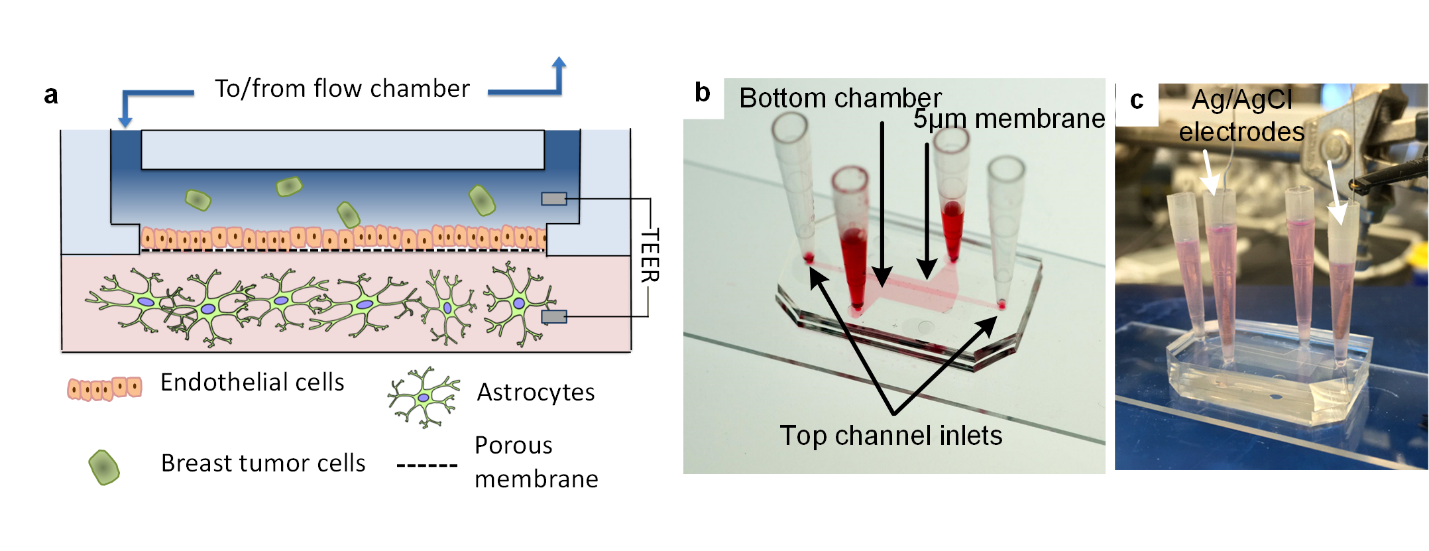
In response to these challenges we have engineered a microfluidic device that recapitulates the human blood-brain barrier niche to study the metastatic process. We first confirm the performance of the device and then measure metastatic potential variation in populations of breast cancer. The device is composed of two chambers (apical vessel and basal brain stroma) separated by a porous membrane. To form a cellular barrier hCMEC cells are seeded onto the apical side of the membrane and cultured to confluence. The basal chamber consists of a collagen ECM and NHA suspension. Cancer cells are introduced into the top chamber and allowed to extravasate into the bottom chamber. Barrier integrity is monitored using trans-epithelia-endothelial-resistance (TEER) and extravasation dynamics are characterized by live-cell microscopy and confocal tomography.
We have applied this new model to compare brain-seeking subclones (MDA-231-BR) of breast cancer cell lines of known whole exome sequence (MDA-231), normal-like cell lines (MCF10A, HME) and brain met patient derived xenografts (PDXs) in terms of their ability to extravasate, migrate and survive in the niche. We characterize their phenotypic and migratory behavior and correlate it to the TEER measurements to establish a metastatic model. Future work, will elucidate the mechanisms that drive specific clones to extravasate using single cell analysis.


