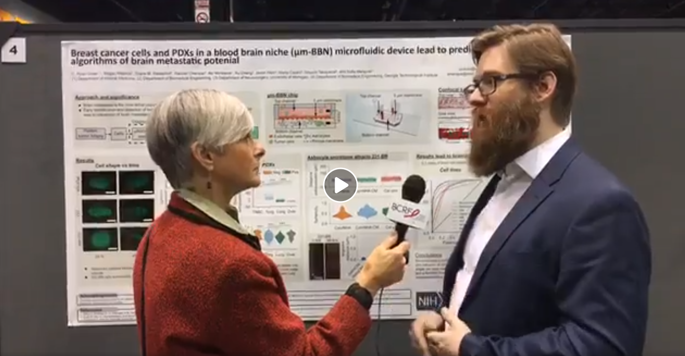
We will be presenting work about a blood brain niche on a chip that have developed to understand clonal variation in breast cancer cells that metastasis to the brain. This is a great event to meet a variety of cancer researchers and understand how biology, materials and microfluidics can be leveraged to solve translational problems.
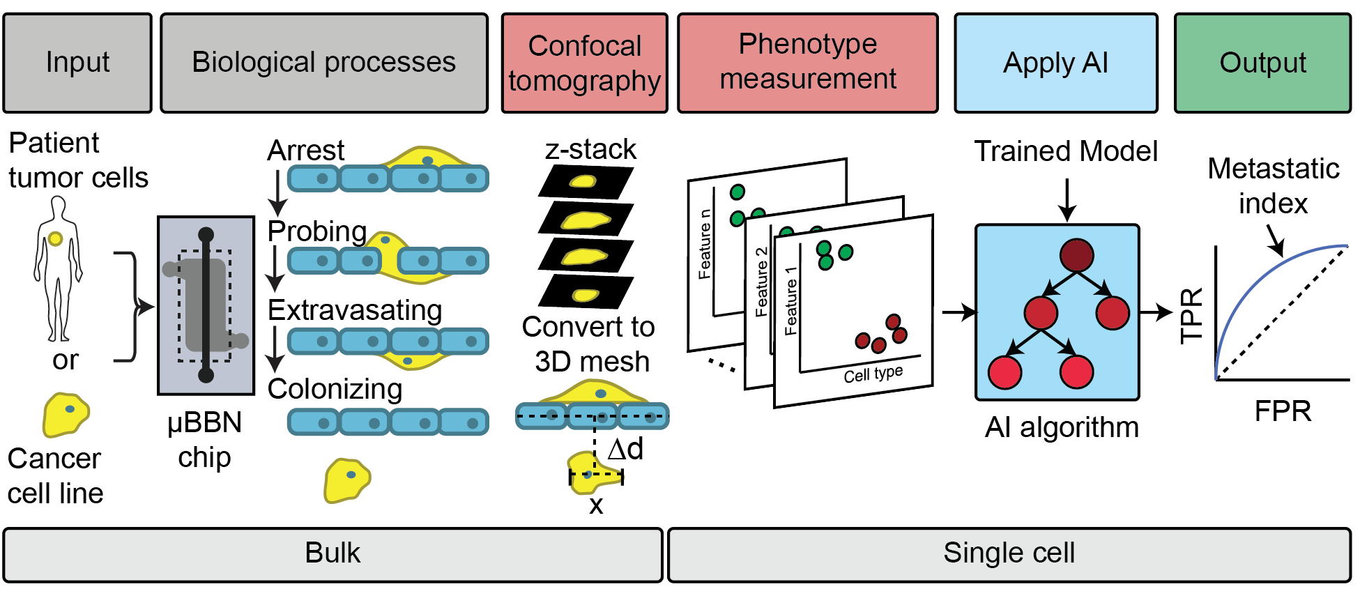
Metastasis from the primary tumor site to the brain is the most lethal complication of advanced cancer. 15% of breast cancers metastasize in the brain with a median survival of 5-14 months depending on the subtype. Therefore, it is critical to identify when a tumor has the clonal potential to metastasize to the brain. Current detection methods and treatment therapies have continued to improve but do not shed light on clonal metastatic potential, moreover, in vivo models do not recapitulate the complexity of the “live” micro-environment. A focus of our group is to develop a microfluidic device that mimics the cellular and physical components of the human blood-brain niche to study the brain metastatic process.
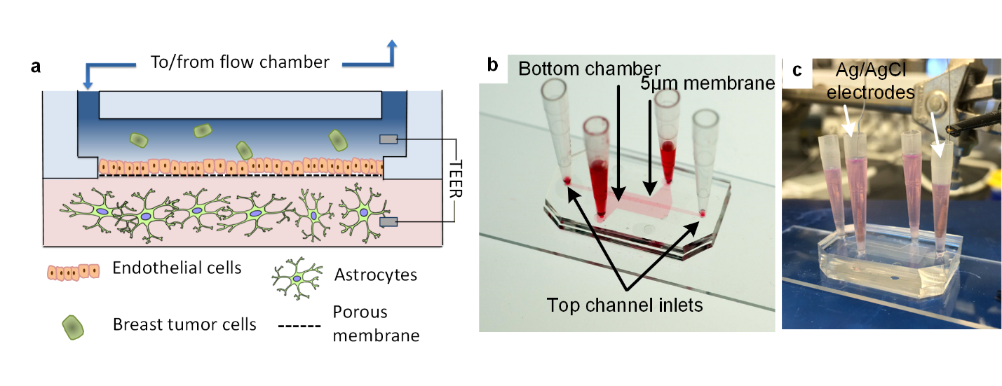
The device is composed of two chambers separated by a porous membrane. The top chamber and apical side of the membrane are seeded with human brain endothelial cells and use flow to mimic shear stress encountered within the vasculature. Cancer cells are introduced into this chamber in which they adhere to and migrate through the endothelium into the bottom chamber. The bottom chamber contains astrocytes suspended in a collagen gel to mimic the brain stroma and provide room for invading cancer cells to colonize and grow. Barrier integrity is monitored using TEER (trans-endothelial electrical resistance), and fluctuates as the tight junctions of the endothelium are compromised by invading cancer cells. This is characterized by IF and tight junction staining. Throughout all time points, from introduction into the flow chamber, adherence to the endothelium, extravasation through the barrier, migration into the stroma, and proliferation the cancer cells can be monitored via both microscopy and TEER.
This work is being done through Dr. Merajver’s lab and is applying this microfluidic blood-brain niche model to compare subclones of breast cancer cell lines in terms of their ability to extravasate, migrate and survive in the niche. We are using this information to characterize their migratory behavior from live-cell microscopy and comparative studies of metastatic markers for µBBN traversing and non-traversing cells when appropriate. We expect this will advance our ability to identify specific cells within heterogeneous populations that have a higher metastatic potential.


